Home

scaramuccia protezione Maledetto collagen staining sviluppo di massimo vantaggio

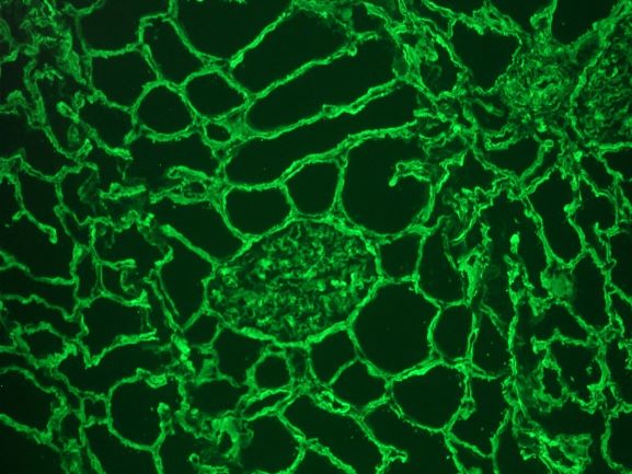
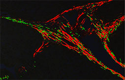
Immunofluorescence staining of extracellular collagen I fibers (green)... |  Download Scientific Diagram
Immunofluorescence staining of extracellular collagen I fibers (green)... | Download Scientific Diagram

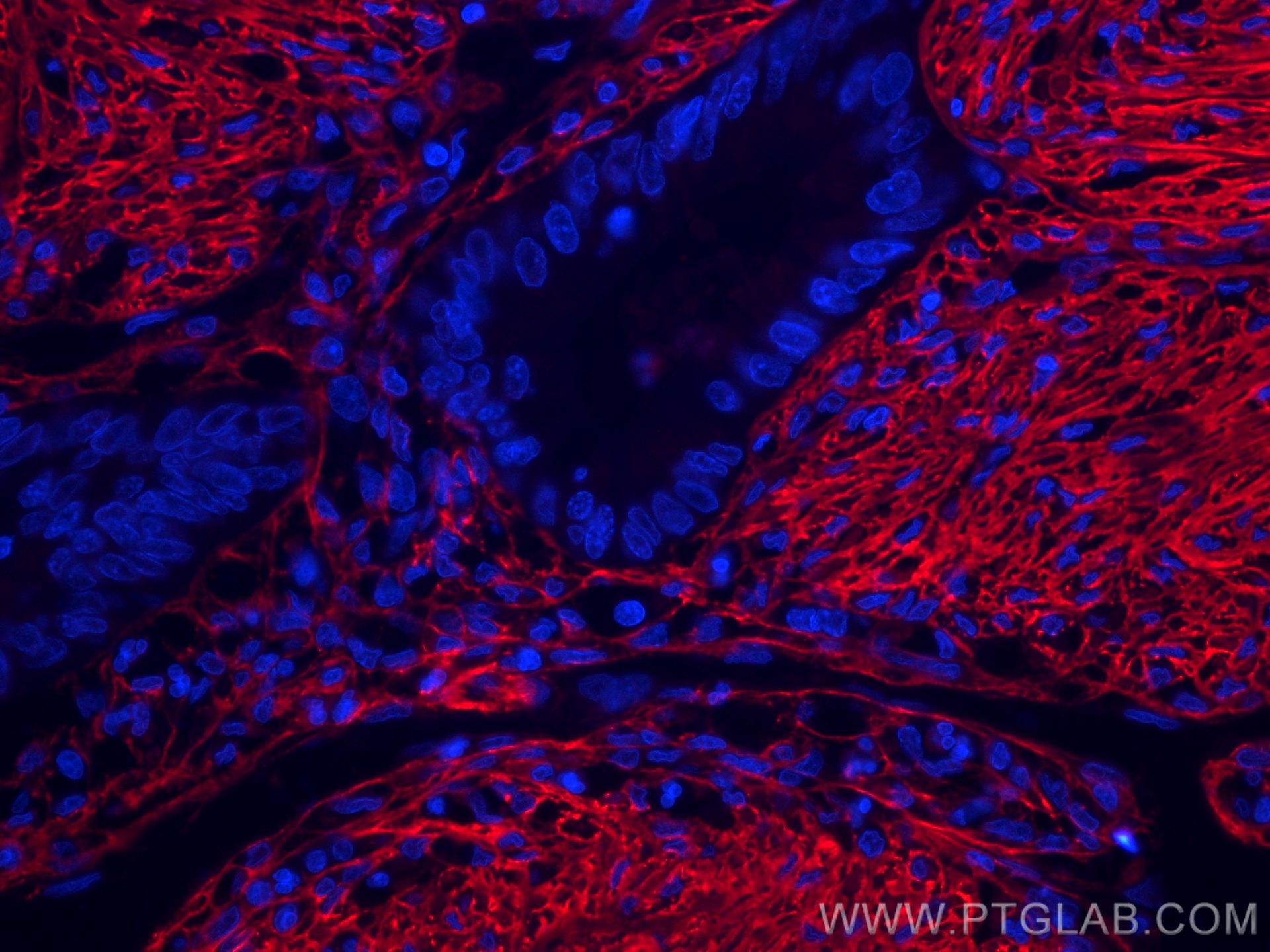
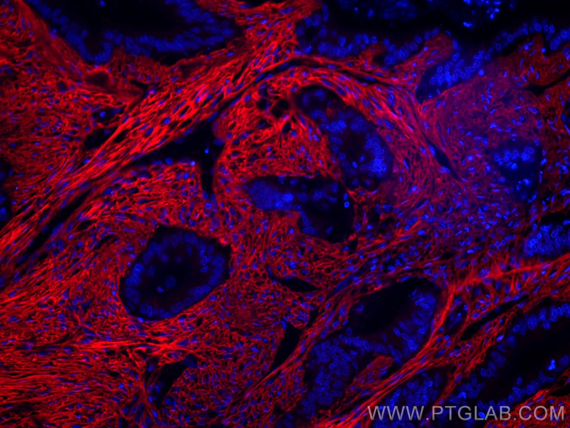
Collagen Type I antibody (CL594-67288) | Proteintech

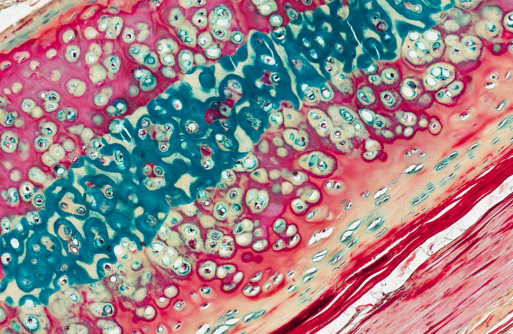
Special Stains | Histology Research Core

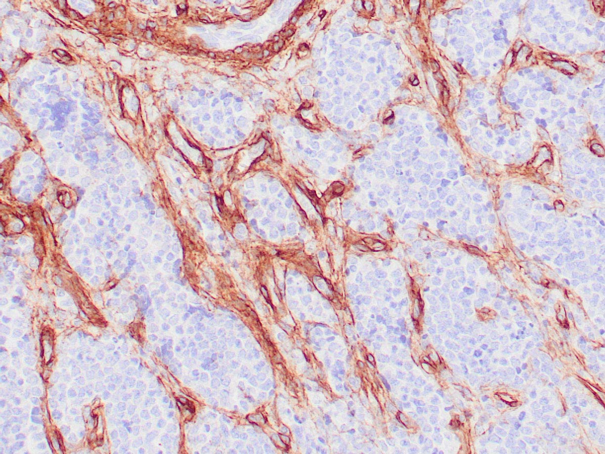
PathPlus™ Collagen IV Polyclonal Antibody Rabbit anti-Human | LSBio

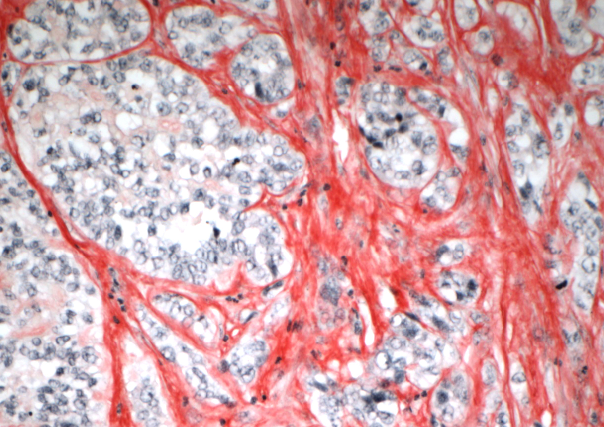
Histochemical Staining of Collagen and Identification of Its Subtypes by  Picrosirius Red Dye in Mouse Reproductive Tissues
Histochemical Staining of Collagen and Identification of Its Subtypes by Picrosirius Red Dye in Mouse Reproductive Tissues

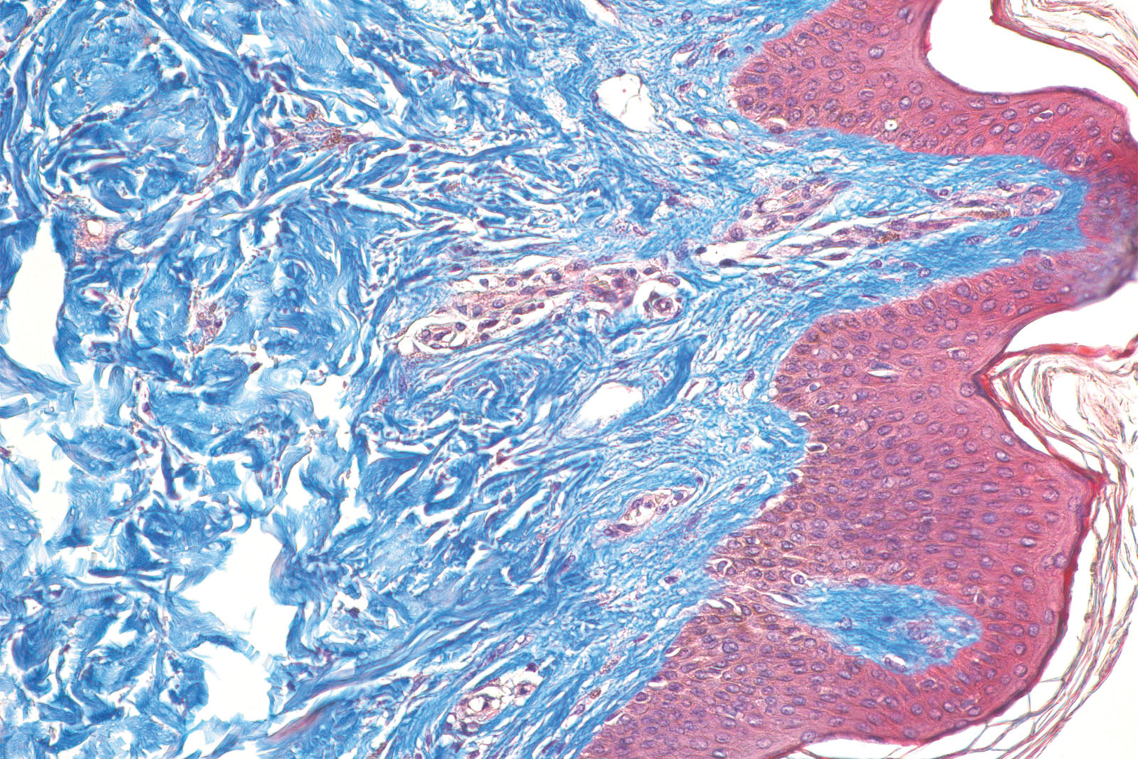
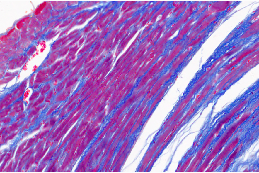
Research SOP: STP FOR MASSON'S TRICHROME STAIN (MT) FOR COLLAGEN

Histochemical Staining of Collagen and Identification of Its Subtypes by  Picrosirius Red Dye in Mouse Reproductive Tissues
Histochemical Staining of Collagen and Identification of Its Subtypes by Picrosirius Red Dye in Mouse Reproductive Tissues
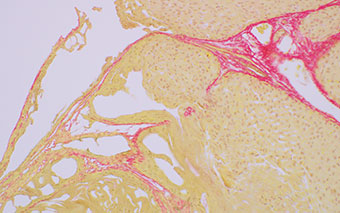
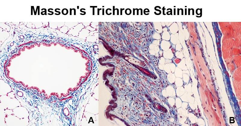
Masson's Trichrome Staining

Identifying Collagen Fiber Types I and III Stained with Picrosirius Red  Using the BX53 Microscope Equipped with Olympus' High Luminosity and High  Color Rendering LED | Olympus LS
Identifying Collagen Fiber Types I and III Stained with Picrosirius Red Using the BX53 Microscope Equipped with Olympus' High Luminosity and High Color Rendering LED | Olympus LS

Staining of human skin with RGB trichrome unveils a proteoglycan-enriched  zone in the hair dermal sheath | bioRxiv
Staining of human skin with RGB trichrome unveils a proteoglycan-enriched zone in the hair dermal sheath | bioRxiv
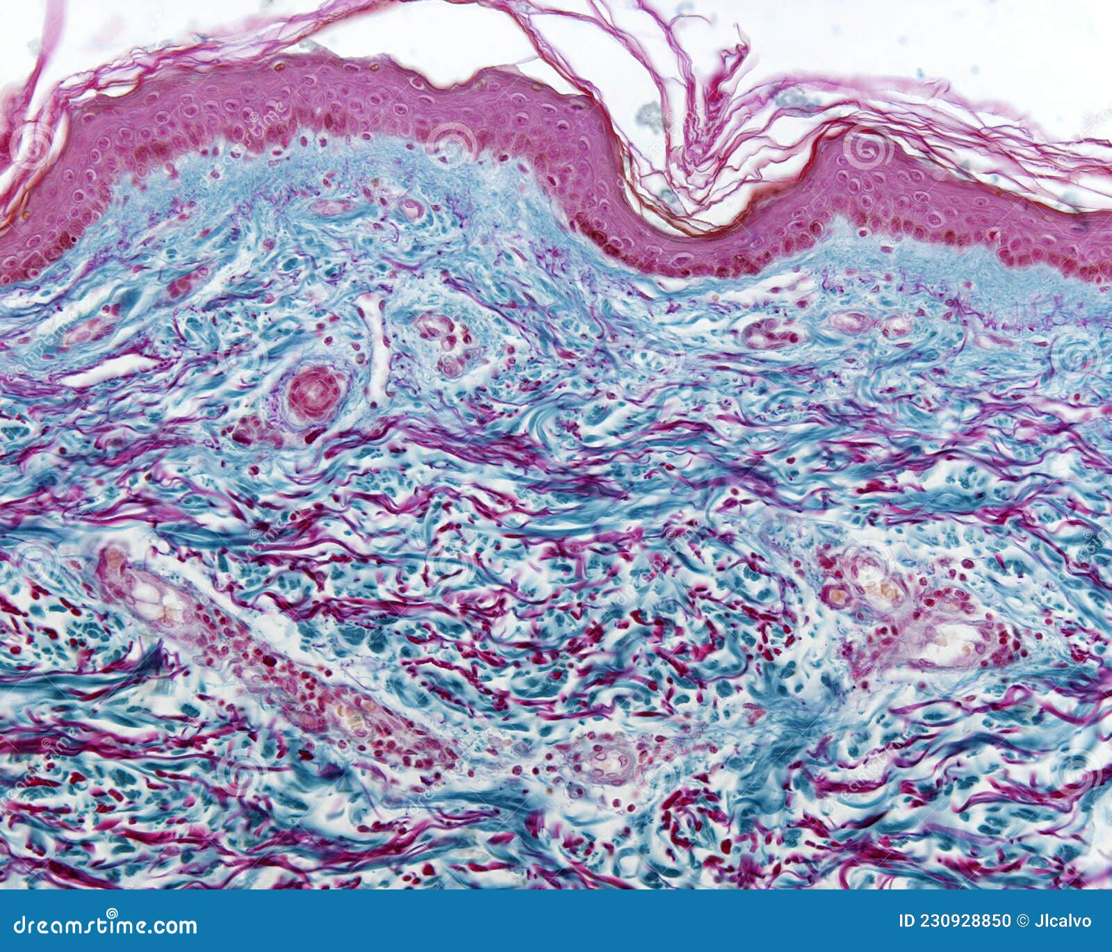
Collagen and Elastic Fibers. Cajal-Gallego Trichrome Stock Photo - Image of  paraffin, cell: 230928850
Collagen and Elastic Fibers. Cajal-Gallego Trichrome Stock Photo - Image of paraffin, cell: 230928850

Collagen Staining in Mouse | Biocompare.com Kit/Reagent Review

VitroView™ Picro-Sirius Red Stain Kit - VitroVivo Biotech

Special Stains for Connective Tissue: Collagen, Reticulin & Elastic Fibers

Collagen IV – Zeta Corporation

Picro Sirius Red Stain Kit (Connective Tissue Stain) (ab150681) | Abcam

Masson's Trichrome staining and collagen type 1 immunoreactivity in the...  | Download Scientific Diagram
Masson's Trichrome staining and collagen type 1 immunoreactivity in the... | Download Scientific Diagram

Masson Trichrome kit - Biognost

Immunohistochemical staining for collagen I, collagen III and... | Download  Scientific Diagram
Immunohistochemical staining for collagen I, collagen III and... | Download Scientific Diagram

Histological staining of collagen in the four different tumors. The... |  Download Scientific Diagram
Histological staining of collagen in the four different tumors. The... | Download Scientific Diagram

Special Stains | Histology Research Core

Immunofluorescence staining of collagen in monolayer cultures of... |  Download Scientific Diagram
Immunofluorescence staining of collagen in monolayer cultures of... | Download Scientific Diagram

Van Gieson's stain - Wikipedia

Collagen Type I Antibody (ABIN5596819)

Determination of collagen content within picrosirius red stained  paraffin-embedded tissue sections using fluorescence microscopy | Semantic  Scholar
Determination of collagen content within picrosirius red stained paraffin-embedded tissue sections using fluorescence microscopy | Semantic Scholar
Identifying Collagen Fiber Types I and III Stained with Picrosirius Red  Using the BX53 Microscope Equipped with Olympus' High Luminosity and High  Color Rendering LED | Olympus LS
Identifying Collagen Fiber Types I and III Stained with Picrosirius Red Using the BX53 Microscope Equipped with Olympus' High Luminosity and High Color Rendering LED | Olympus LS

Comparison of Picrosirius Red Staining With Second Harmonic Generation  Imaging for the Quantification of Clinically Relevant Collagen Fiber  Features in Histopathology Samples - Cole R. Drifka, Agnes G. Loeffler,  Kara Mathewson, Guneet
Comparison of Picrosirius Red Staining With Second Harmonic Generation Imaging for the Quantification of Clinically Relevant Collagen Fiber Features in Histopathology Samples - Cole R. Drifka, Agnes G. Loeffler, Kara Mathewson, Guneet

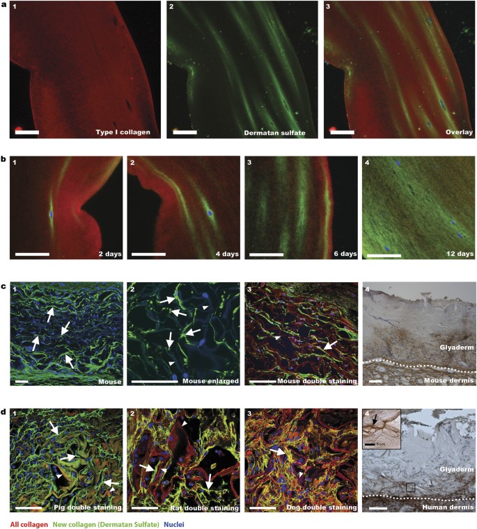
Visualisation of newly synthesised collagen in vitro and in vivo |  Scientific Reports
Visualisation of newly synthesised collagen in vitro and in vivo | Scientific Reports

Collagen Type I antibody (CL594-67288) | Proteintech